Anatomy Diagram Rib Area - Anatomy Diagram Rib Area - Rib Anatomy at University of ... / Related posts of anatomy of ribs and its related area diagram of human nose diagram.
Anatomy Diagram Rib Area - Anatomy Diagram Rib Area - Rib Anatomy at University of ... / Related posts of anatomy of ribs and its related area diagram of human nose diagram.. The rib cage surrounds the lungs and the heart, serving as an important means of bony protection for these vital organs. Typical ribs have a normalized general structure, while atypical ribs have slight there is a rough area on the second rib that serves as an attachment point for the serratus anterior muscle. More than half of spinal cord injuries occur in the cervical area, a third occur in the thoracic area, and. They extend from the lateral border of the costal grooves to the superior margins of the ribs below. Pain+left+side+under+ribs | intro to anatomy 6:
The rib cage surrounds the lungs and the heart, serving as an important means of bony protection for these vital organs. More than half of spinal cord injuries occur in the cervical area, a third occur in the thoracic area, and. Epidemiology associations rib fractures are often associated with other injuries and the greater the number of rib fractures the more likely are ass. Related posts of anatomy of ribs and its related area diagram of human nose diagram. Rib number 10 is atypical because its head.
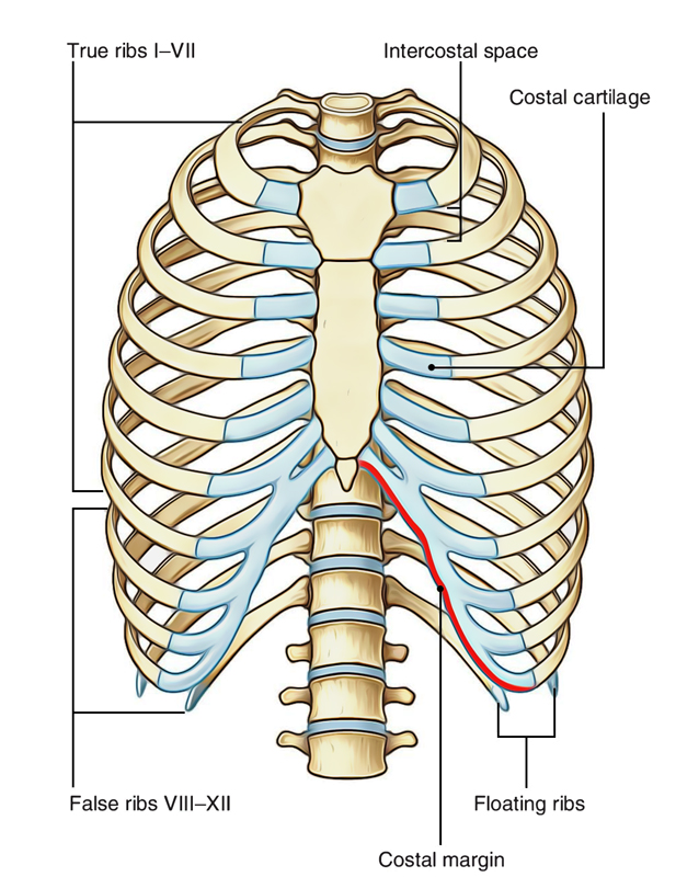
ribs anatomy from www.earthslab.com Rib number 10 is atypical because its head. The rib cage surrounds the lungs and the heart, serving as an important means of bony protection for these vital organs. The rib cage, shaped in a mild cone shape and more flexible than most bone sets, is made up of varying elements such as the thoracic vertebra, 12 equally paired ribs, costal cartilage, and held together anteriorly by the sternum. They articulate with the vertebral column posteriorly, and terminate anteriorly as cartilage (known as costal cartilage). The ribs are the skeletal protection for the lungs and the chest cavity. Its small branches supply blood to the ribs and some chest structures. The ribs and rib muscles expand and contract with normal breathing. Human breathing, lung capacities, and breathing cycles.
Learn everything about the ribs with our articles, video tutorials, quizzes, and labeled diagrams there are eleven pairs of external intercostal muscles and these are the most superficial in the area.
The current morbidity of rib plating is due to the size of the incision required to perform an open procedure. Start studying anatomy of the rib. These are large areas of the cerebral cortex that receive sensory input from multiple. The skull and rib cage. Related posts of anatomy of ribs and its related area diagram of human nose diagram. The ribs are a set of twelve paired bones which form the protective 'cage' of the thorax. As part of the bony thorax, the ribs protect the internal thoracic organs. Human brain functional infographic diagram. The distribution of air sacs and the functioning of the avian lung. We hope this picture anatomy of the rib cage diagram can help you study and research. Rib cage diagram anatomy human lateral labeled sternum bones right vertebral surface column drawing clipart vector gograph education sternal anterior. The ribs and rib muscles expand and contract with normal breathing. The ribs are the skeletal protection for the lungs and the chest cavity.
As part of the bony thorax, the ribs protect the internal thoracic organs. Just like in the manubrium. It also includes some the facets and demifacets devoted to rib articulation demonstrate the main function of the thoracic spine. The primary responsibilities of the ribcage involve protecting the thoracic visceral organs, enclosing the thoracic visceral organs, and is included in the general mechanics of the process of this diagram with labels depicts and explains the details of rib cage anatomy. See more ideas about human anatomy, anatomy, anatomy reference.
Are The Kidneys Located Inside Of The Rib Cage - Kidney ... from www.anatomylibrary99.com The rib cage, shaped in a mild cone shape and more flexible than most bone sets, is made up of varying elements such as the thoracic vertebra, 12 equally paired ribs, costal cartilage, and held together anteriorly by the sternum. Webmd's aorta anatomy page provides a detailed image and definition of the aorta. Learn vocabulary, terms and more with flashcards, games and other study tools. All are attached at the back to the thoracic vertebrae and are numbered from 112 according to the vertebrae they attach to. Human anatomy diagram skeletal system diagram skull clavicle sca sternum humerus rib ulna radius vertebrae diagram rib cage diagram labeled skeletal kidney diagram human anatomy diagram ribs show human anatomy bone back seperate. Human anatomy abdominal organs abdominal diagram with ribs anatomy. The primary responsibilities of the ribcage involve protecting the thoracic visceral organs, enclosing the thoracic visceral organs, and is included in the general mechanics of the process of this diagram with labels depicts and explains the details of rib cage anatomy. Learn everything about the ribs with our articles, video tutorials, quizzes, and labeled diagrams there are eleven pairs of external intercostal muscles and these are the most superficial in the area.
There are two types of ribs, namely typical and atypical.
The current morbidity of rib plating is due to the size of the incision required to perform an open procedure. In most tetrapods, ribs surround the chest, enabling the lungs to expand and thus facilitate breathing by expanding the chest cavity. Click the image to watch the anatomy of the rib cage video. The ribs are the skeletal protection for the lungs and the chest cavity. Pain+left+side+under+ribs | intro to anatomy 6: Human breathing, lung capacities, and breathing cycles. Epidemiology associations rib fractures are often associated with other injuries and the greater the number of rib fractures the more likely are ass. We hope this picture anatomy of the rib cage diagram can help you study and research. Learn everything about the ribs with our articles, video tutorials, quizzes, and labeled diagrams there are eleven pairs of external intercostal muscles and these are the most superficial in the area. Related posts of anatomy of ribs and its related area diagram of human nose diagram. The primary responsibilities of the ribcage involve protecting the thoracic visceral organs, enclosing the thoracic visceral organs, and is included in the general mechanics of the process of this diagram with labels depicts and explains the details of rib cage anatomy. Human brain functional infographic diagram. It has a roughened area on its upper surface, from which the serratus anterior muscle originates.
Overlying flaps projecting off the ribs called uncinate at the end of the digestive tract is the cloaca, a holding area for wastes and products from the figure 9. Learn about its function and location as well as conditions that affect the aorta. The rib cage, shaped in a mild cone shape and more flexible than most bone sets, is made up of varying elements such as the thoracic vertebra, 12 equally paired ribs, costal cartilage, and held together anteriorly by the sternum. They also have a role in. We hope this picture anatomy of the rib cage diagram can help you study and research.
Chest Anatomy Diagram - Cheat Dumper from i.pinimg.com The first seven are connected behind with the vertebral column and in front. Rib number 10 is atypical because its head. Click the image to watch the anatomy of the rib cage video. Includes images, video, and free quiz. Its small branches supply blood to the ribs and some chest structures. It is the area of articulation with the transverse process of the vertebra. Just like in the manubrium. Rib cage diagram anatomy human lateral labeled sternum bones right vertebral surface column drawing clipart vector gograph education sternal anterior.
Human breathing, lung capacities, and breathing cycles.
Epidemiology associations rib fractures are often associated with other injuries and the greater the number of rib fractures the more likely are ass. The ribs are the skeletal protection for the lungs and the chest cavity. Rib number 10 is atypical because its head. Rib cage anatomy britannica com. There are two types of ribs, namely typical and atypical. Click the image to watch the anatomy of the rib cage video. Includes images, video, and free quiz. Human brain functional infographic diagram. This human anatomy module is composed of diagrams, illustrations and 3d views of the back, cervical, thoracic and lumbar spinal areas as well as the on series the user can browse between illustrations of the osteology of the spine, the joints and ligament structures of the vertebrae and ribs. Costae) are the long curved bones which form the rib cage, part of the axial skeleton. Start studying anatomy of the rib. Learn vocabulary, terms and more with flashcards, games and other study tools. Learn everything about the ribs with our articles, video tutorials, quizzes, and labeled diagrams there are eleven pairs of external intercostal muscles and these are the most superficial in the area.


Posting Komentar
0 Komentar